Robotic Umbilical Hernia Surgery
An umbilical hernia occurs when part of the intestine protrudes through an opening in the abdominal wall at the belly button, and it is usually harmless. This condition is most commonly seen in infants. During fetal development, the umbilical cord, which connects the baby to the mother, passes through a small opening in the abdominal wall.
This opening is expected to close on its own shortly after birth. If it does not close, fatty tissue or a portion of the intestine can protrude through the opening, leading to an umbilical hernia. A bulge at the belly button, especially when the baby cries, may be a sign of an umbilical hernia.
Diagnosis and Treatment Methods of Umbilical Hernia
An umbilical hernia occurs when a portion of the intestine protrudes through the belly button opening in the abdominal muscles and is usually harmless. This condition is most commonly seen in infants. During fetal development, the umbilical cord that connects the baby to the mother passes through a small opening in the abdominal wall. This opening is expected to close shortly after birth. If it doesn’t, fatty tissue or a part of the intestine can protrude through the opening, leading to an umbilical hernia. A bulge at the belly button may be observed when the baby cries. In most cases, it heals on its own by the age of 1–2, but in some cases, the healing process may take longer.
In adults, excessive abdominal pressure can cause fatty tissue or a portion of the intestine to push through a weakened area in the abdominal muscles, resulting in an umbilical hernia. Factors such as obesity, heavy lifting, previous surgeries, and a family history of hernias can trigger umbilical hernias in adults.
Diagnosis:
Umbilical hernias can be easily diagnosed through physical examination. In some cases, imaging techniques such as ultrasound or CT scans may be used to assess potential complications. In infants, the hernia is expected to disappear on its own by the age of 4. However, umbilical hernias in children over 4 years of age or in adults may require surgical intervention.
Treatment Methods:
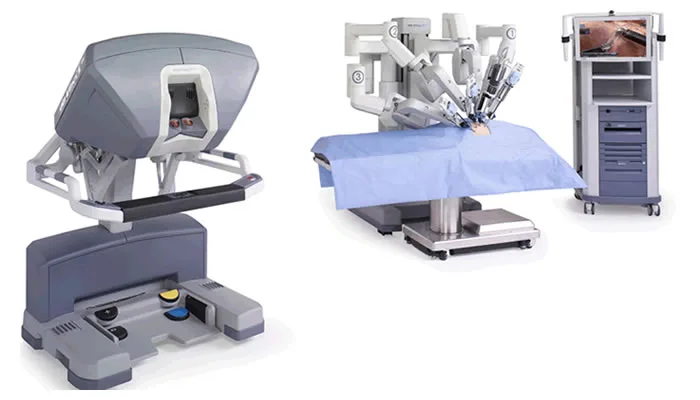
Robotic Surgery (Robot-Assisted Minimally Invasive Surgery):
Robotic surgery allows the surgeon, positioned a few steps away from the patient, to perform the operation under high-resolution 3D visualization provided by the robot’s specialized camera. This method is performed through a few small incisions, which speeds up the patient’s recovery process. The surgeon’s hand movements are instantly transmitted to robotic instruments, allowing for a more precise and accurate operation. The small incisions promote quicker postoperative healing and enable patients to return to their daily activities sooner.
Open Surgery:
Open surgery can be performed under local or general anesthesia. In this method, the surgeon makes an incision in the abdominal area and pushes the protruding tissue back into the abdominal cavity. The weakened area is then sutured and closed. Since open surgery involves larger incisions, the recovery process may take longer.
Laparoscopic Surgery (Minimally Invasive Surgery):
Laparoscopic surgery is performed through several small incisions in the abdominal area. During the procedure, the abdomen is inflated with gas to enlarge the surgical area. A small camera is inserted through one of the incisions, and the surgeon performs the operation using the visual guidance. Laparoscopic surgery is a minimally invasive method that accelerates the patient’s recovery and allows for a quicker return to normal activities.
TREATMENTS